ChimeraX docs icon

Toolbar: Medical Image

For convenience in working with medical image data, the Medical Image tab of the Toolbar brings together several related icons, many of which also appear in other tabs. See also: ChimeraX DICOM Reference, DICOM in VR tutorial, DICOM segmentation tutorial, ChimeraX for Medical Image Analysis

File
open DICOM same as Menu: File... Open DICOM Folder

Home
toggle main window between 3D graphics and recent files (Rapid Access) same as clicking bottom right icons  

Home
save snapshot of main window  save ~/Desktop/imageN.png supersample 3

(N = 1,2, ...)

Display

Map
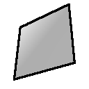
single Z-plane at the center of the current region; assign right mouse button to move planes volume planes z style image imageMode "full region"
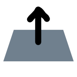
+
mousemode right "move planes"

Map
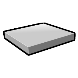
tilted-slab image display, with initial slab in the middle of the data, 10 planes thick and perpendicular to the data Z axis; assign right mouse button to rotate slab volume with style image and imageMode "tilted slab"

(exact command depends on the data) +
mousemode right "rotate slab"

Map
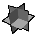
XYZ orthoplanes at the center of the full data; assign right mouse button to move planes ; see also ui view volume to set orthoplanes with style image region all
(exact command depends on the data) +
mousemode right "move planes"

Map
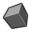
full region; assign right mouse button to crop volume volume region all imageMode "full region"
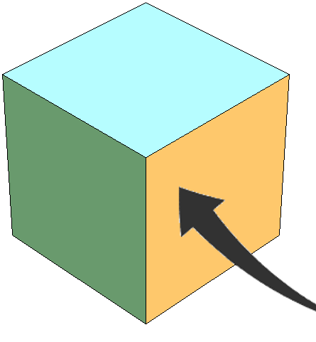
+
mousemode right "crop volume"

Map
toggle outline box volume with the showOutlineBox option
airways preset for image display volume appearance airways
chest preset for image display volume appearance chest
brain preset for image display volume appearance brain
initial thresholds preset for image display, e.g., for single planes volume appearance initial

Graphics
view all, standard orientation view orient

Map
step 1, full resolution of data volume step 1

Map
step 2, subsampled at every other point volume step 2
2D-XYZ projection for image display volume projectionMode 2d-xyz
3D projection for image display volume projectionMode 3d
raycasting for image display volume projectionMode rays

Mouse Modes in the Medical Image Toolbar

The last (rightmost) icons assign the function or “mouse mode” of:

icon mouse mode function

Right Mouse
crop volume show volume data outline box and adjust region size by dragging any face; see also volume

Right Mouse
move planes show single plane of clicked volume data if full region shown initially; move plane or slab along its axis to show a different section

Right Mouse
rotate slab activate tilted slab slicing of volume data (image style only), rotate the slab to adjust the slicing angle; the center of rotation from clicking over the slab is the center of the slab, and from clicking elsewhere is the center of the volume box;
assigned button + Shift translates the slab along its axis; in VR, this mode both rotates and translates the slab

Right Mouse
tape measure click to anchor distance measurement, drag to desired endpoint; click in empty space to “erase” (details...)

Right Mouse
windowing adjust volume data image display thresholds collectively: vertical motion adjusts window level (moves all thresholds in parallel to higher or lower values), horizontal motion adjusts window width (moves thresholds symmetrically farther apart or closer together), and the dominant type of motion wins out; for adjusting multiple windows within a single histogram, see details
(multiple) toggle mouse button functions: assign them as specified in the Mouse (3D) settings of the Segmentations tool, or restore the previous functions; see also segmentations mouseModes
(multiple) toggle VR hand-controller functions: assign them as specified in the VR Controller settings of the Segmentations tool, or restore the previous functions; see also segmentations handModes

We gratefully acknowledge support from the NIAID for ChimeraX development related to medical image analysis.


UCSF Resource for Biocomputing, Visualization, and Informatics / July 2024