 |
|
Bacteriorhodopsin
 JPEG version (133KB), TIFF version (818KB)
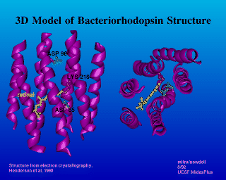
This image was rendering using the MidasPlus delegate program Cartoon. It shows the structure of Bacterorhodopsin, and the location of retinal. The colored background was achieved using an external file with the background color in it. The file might look like the following.
The file ``color.file'':
background 0.0 0.0 1.0To use this file, within MidasPlus, the command:
ribbon -c color.fileSee the Cartoon manual page for more details.
The labels were created with Ilabel, another MidasPlus program.
There is a new program called Ribbonjr, which also draws ribbons through proteins. It is more up to date, and can also make solid ribbons that can be interactively rotated using the SceneViewer utility program on the Silicon Graphics machine. Ribbonjr is now the preferred program to Cartoon, so please see the manual pages for Ribbonjr.
The structure of Bacteriorhodopsin was solved by Henderson, et. al., 1990. Image created by Julie Newdoll and Alok Mitra. ©2004 The Regents, University of California; all rights reserved.